How we use PET-CT scans to find more cancer faster than ever
Share this
PETCT-scan-benefits-blog-desktop

PET-CT  (positron emission tomography and computed tomography) often is referred to as the gold standard of cancer imaging. This tool brings together two powerful technologies to give us advanced information about whether a patient has cancer and, if so, where the cancer is located in the body.

Your doctor may order a PET-CT scan after noticing something unusual during another test, such as an X-ray or MRI (magnetic resonance imaging) scan, or they may order one after you’ve reported unusual symptoms leading to a physical examination. For example, I’ve seen patients who visited their regular doctor for a little tickle in their throat, and their doctor saw a small, worrisome-looking growth during an exam, prompting the need for a biopsy and subsequent PET-CT scan to look for other sites of tumor. Sometimes it turns out that the tickle was caused by cancer within a patient's neck and the patient may also have disease elsewhere, for example in the chest.

A combined PET-CT scan can give us more information about a tumor than a PET or a CT alone. It combines two of the most important features of any lesion that is worrisome for cancer: its anatomy and its metabolism (presence of live cells). A true example of how these two features can be combined and then distinguished from one another is when you see a large mass or tumor on a CT scan, but a PET scan might show that only the inner part of the mass is cancerous. This allows us to focus radiation, for example, in just the diseased area of the mass, reducing damage to healthy surrounding tissue. Our hybrid scanner combines (fuses) these images to give us the whole picture, which improves the accuracy of the diagnosis and can reduce the number of scans a patient needs.

LISTEN: Dr. Garcia discusses the benefits of PET-CT scans in the Medical Intel podcast.

How a PET-CT scan works

The CT portion works just like a regular CT scan, using multiple X-ray images to provide a clearer, more detailed image than a single X-ray picture. A CT scan shows anatomic images of tumors or masses, but occasionally it may fail to detect tissue damage or changes that occur in the early stages of cancer. That’s where the PET portion of the scan becomes important.

PET detects the presence of positrons, which are particles smaller than an atom that have a positive electrical charge. Positrons are emitted by radioactive materials as they’re broken down. All cells use glucose, or sugar, for energy. We use a radioactive form of glucose called Fluorodeoxyglucose (FDG) attached to a radioactive substance called fluorine-18. As the cells absorb the sugar as an energy source, the fluorine-18 will break down, and give off positrons and gamma rays that will shine or glow, if you will, on imaging.

Cancer cells tend to be more active than normal cells because they are growing and multiplying faster than normal, so they take in more sugar and give off more positrons and gamma rays. This causes them to glow brighter than normal cells on a PET-CT scan. Some of the cancers that are most easily detected on a PET-CT scan are breast cancer, lung cancer and lymphoma.

#Breastcancer, #lungcancer and #lymphoma are a few of the cancers that a #PETCT scan can detect most easily. bit.ly/2Dssakc via @MedStarWHC
Click to Tweet

Cancer is like a living disease that eats and grows. We use PET-CT to monitor the activity of the disease and watch whether the number of cancer cells is decreasing, along with the shine of the imaging, which is a good indication that the patient is beating the disease.

While PET scanning does apply a higher dose of radiation to the body at once than CT imaging alone or multiple X-rays, the amount of information your doctor can glean from the fused imaging outweighs this risk for most patients. PET-CT scanning is safe for nearly every patient. In fact, we wish we could use PET-CT scanning for more diseases because of the crisp images and ability to track diseased cells.

What to expect during your scan

If your doctor has ordered a PET-CT scan for you, we’ll ask you to not eat or drink anything except water for six hours before the test . In many instances, you will also be asked to not exercise 24 hours before your exam. And since we would want the radioactive sugar to be the first energy your body receives, you should also avoid consuming sugars 24 hours before your exam, so your cells will latch onto it quickly. We’ll give you the sugar in an IV injection, and then you’ll sit in a warm, quiet room for 40 to 60 minutes so you can relax and let the sugar absorb into your cells without any stimulus to your body.

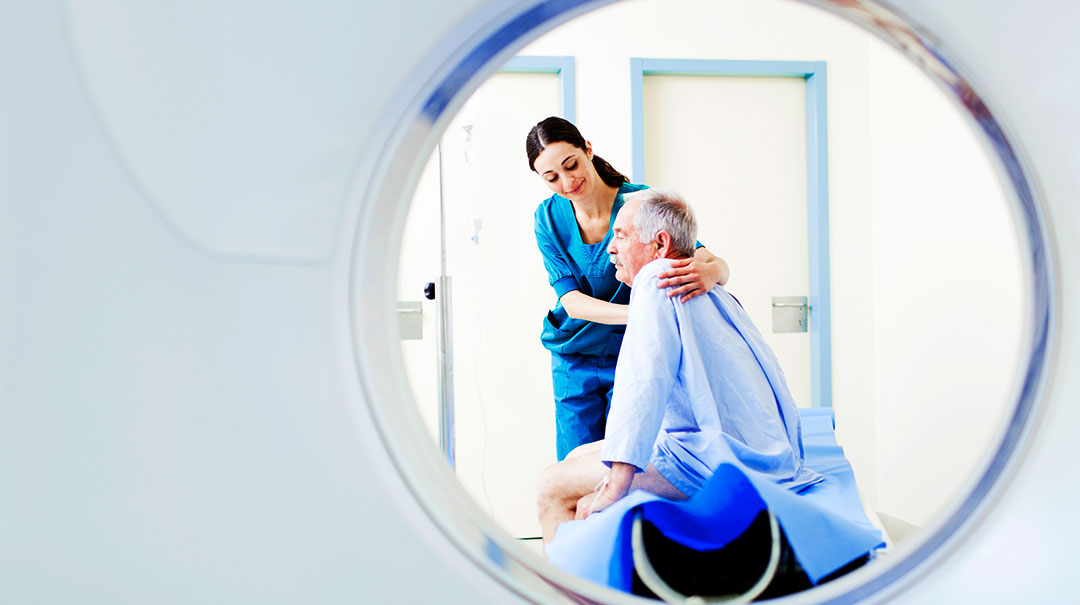
The scan itself usually takes 20 to 30 minutes. You will lie on a table that will pass through the PET-CT scanner several times as the machine takes pictures from your eyes to about the middle of your thighs. Once that’s done, you can go home. We’ll review your images to interpret what they show, and then we’ll report our results to your doctor.

If the PET-CT scan finds cancer, your doctor can assign a stage to the cancer based on the images of its size and whether it has spread to other organs and/or lymph nodes. Smaller, lower-stage cancers that have not spread often can be removed with surgery or destroyed through radiation therapy. Larger, more advanced cancer that has spread may require whole-body treatment, such as chemotherapy.

As treatment progresses, we use follow-up PET-CT scans to monitor your response. If the treatment is working, we should see smaller and fewer bright areas on the scan, which indicates that the cancerous cells are dying off. If the treatment isn’t working, we’ll see more bright areas and areas that are brighter than past scans, which means the cancer is growing. That tells the oncologist that a different treatment might be needed.

Nothing makes me happier than to compare an old scan of a patient that showed lots of lit-up, cancerous areas, to a new one that’s a blank canvas, showing that the cancer is gone after treatment. With the latest advances in PET-CT technology, I’m confident we’ll be able to share these happy days with even more patients and their cancer doctors in the years to come.

Stay up to date and subscribe to our blog

Latest blogs