If you are experiencing a medical emergency, please call 911 or seek care at an emergency room.
In 2006, the latest technology available for PET-CT scans was the time-of-flight scanner. At the time, this scanner compared to older PET models was like comparing old, heavy TVs of the 1980s to the first generation of flat screens. We made the jump to the time-of-flight scanner when it first became available, and we were the only center in our area to have the technology for many years.
But just like TV technology, PET-CT scanners have continued to get better since the time-of-flight scanner. Today’s digital PET-CT scanners provide a huge improvement in image quality.
We’re now able to detect smaller cancer lesions we might not have seen before. MedStar Washington Hospital Center was one of the first in the region to have the most advanced digital scanning unit in the world. Think of it as the ultra-high-definition, curved-screen TV of the imaging world—it’s a night-and-day difference over what we had previously.
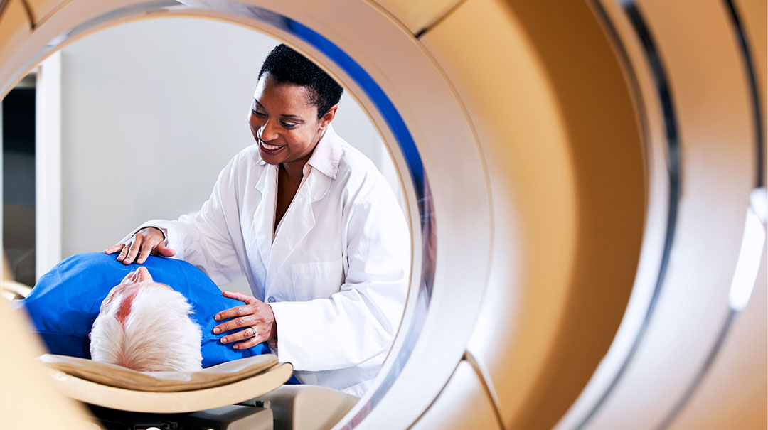
PET-CT scanning is the gold standard of cancer imaging today. It’s approved for detecting and monitoring virtually every type of cancer. The advanced digital scanners offer many benefits over time-of-flight scans.
The patient gets:
Half the time in the scanning machine
Half the dose of radiation
Images that are 10 times crisper
The digital scanner makes it possible for us to better detect cancer lesions—even smaller ones that we might not have been able to see before. Detecting these lesions early means doctors may be able to treat a patient’s cancer sooner and more effectively.
LISTEN: Dr. Garcia discusses advances in PET-CT technology in the Medical Intel podcast.
How PET-CT scanning helps us detect tricky thyroid tumors
Our expertise with PET-CT scanning also can benefit some patients with thyroid cancer. Thyroid cells sometimes can lose the ability to absorb iodine in certain types of thyroid cancer. Normally, thyroid cancer cells are easy to track with radioactive iodine in a test called radioiodine imaging because of their unique ability to absorb iodine, which they use to make thyroid hormone. If that ability is gone, we have to turn to PET-CT scanning.
The PET-CT test uses a form of radioactive sugar, rather than regular iodine, to locate cancer cells. Normally, patients are given a radioactive iodine pill, and the body treats it like regular iodine, sending it anywhere in the body where there is thyroid tissue. Wherever the cancer cells have spread, the cancer cells will act like thyroid cells, just growing elsewhere in the body; for example, the lungs or the bones. The radioactive iodine is chemically altered to get “stuck” in the cell, once absorbed, and not made into thyroid hormone, so we can spot it on imaging.
Even if the cells are no longer able to take up iodine, the cells might still be able to take up glucose, which cancer cells use for energy. We can use PET-CT scanning to watch the metabolic reaction of cells throughout the body to spot cancers and determine the best course of care for individual patients. Basically, we trick the body into doing what it normally does, and we use that information to treat patients’ cancer.
Sharing our PET-CT knowledge with future doctors
The field of nuclear medicine grows and evolves all the time, and doctors need to grow along with it. We’re involved in that process, by helping to organize the continuing medical education courses all nuclear medicine doctors have to take, to stay up-to-date with the latest scientific advances. In this way, we’re shaping the direction of nuclear medicine. Much of our research surrounds determining how much radiation is delivered into lesions, so we can begin to tailor those levels to specific patients’ needs.
In addition, we worked extensively with the second installment of the National Oncologic PET Registry (NOPR) in a six-year process to analyze the use of PET-CT scanning for cancers that appear on the bone, whether they’ve started there or spread from other areas. We were one of the major cancer treatment centers that helped collect this sort of information. This effort leads leads developing of guidelines other doctors and hospitals can use, to direct them when testing for bone cancers, or cancers that tend to spread to the bones when they reach more advanced stages.
We worked with the first installment of NOPR on a previous six-year process of establishing when to use PET-CT scans to locate virtually any type of cancer in the body. I am proud to say that now almost every cancer in any stage is approved to be examined with a PET-CT scan, which was an enormous accomplishment and a nationwide team effort.
Staying ahead of the latest uses for PET-CT and the technology that makes these developments possible is critical for us, as nuclear medicine doctors. We’re able to provide better care and outcomes for the patients of today, and we’re helping prepare future doctors to provide that care to patients for many years to come.